Anatomical Models about Femoral Fracture and Hip Osteoarthritis
This hip fracture model is ideal for orthopaedic departments, physio and GP clinics.
Notify me when back in stock
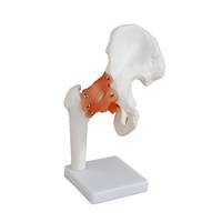
It shows the right hip joint of an elderly person in half natural size.
In addition to the external anatomy of the hip joint, a frontal section through the femoral neck is shown in relief on the base. The hip joint model shows the femoral fractures that occur most commonly in practice as well as typical wear and tear symptoms of the hip joint.
The following fractures are shown on the hip joint:
- Medial femoral neck fracture
- Lateral femoral neck fracture
- Fracture through the trochanteric region
- Fracture below the trochanters
- Femoral shaft fracture
- Femoral head fracture
- Fracture of the greater trochanter
(A88)
- Medial femoral neck fracture
- Lateral femoral neck fracture
- Fracture through the trochanteric region
- Fracture below the trochanters
- Femoral shaft fracture
- Femoral head fracture
- Fracture of the greater trochanter
Weight: 0.4 kg
Dimensions: 14 x 10 x 22 cm
| SKU | A88 |
| Barcode # | 4053083001521 |
| Brand | 3B Scientific |
| Shipping Weight | 0.2000kg |
| Shipping Width | 0.140m |
| Shipping Height | 0.200m |
| Shipping Length | 0.100m |
Warranty: 3 years
Be The First To Review This Product!
Help other Device Technologies Pty Ltd users shop smarter by writing reviews for products you have purchased.


